Appendix Cancer

Appendix cancer, or appendiceal cancer, arises when the cells of the appendix mutate and proliferate uncontrollably, forming a tumor. It is often discovered during appendicitis surgery or unrelated imaging tests. Smaller tumors have a lower tendency to spread, while larger ones typically require more aggressive treatment.

Appendix Cancer Abridged

What is Appendix Cancer?

Appendix cancer is a rare form of cancer that occurs when cells in the appendix undergo mutations, leading to uncontrolled growth. The appendix is a small pouch in the digestive system located at the junction of the large and small intestines. Its function is not fully understood, but it may play a minor role in the immune system’s functions, or it could be an entirely redundant vestigial organ.

Types of Appendix Cancer

Appendiceal Adenocarcinoma: Originates in the glandular tissue of the appendix. 

Variants include:

  • Mucinous Adenocarcinoma: Produces mucin and typically remains within the abdomen.
  • Colonic-Type Adenocarcinoma: Resembles colon cancer, emerging at the appendix base.
  • Signet Ring Cell Adenocarcinoma: A rare form characterized by cells with a signet ring appearance.
  • Goblet Cell Adenocarcinoma: Combines features of adenocarcinoma and neuroendocrine tumors.

Appendiceal Neuroendocrine Tumors: Generally slow-growing tumors that affect neuroendocrine cells, which regulate hormone release in response to nerve signals.

Prevalence

Appendix cancer is extremely rare, affecting about 1 to 2 people per million annually in the United States.

Symptoms of Appendix Cancer

Appendix cancer might not show the following symptoms until it spreads:

  • Appendicitis-like pain
  • Abdominal bloating or fluid buildup
  • Increased waist size
  • Abdominal or pelvic pain
  • Changes in bowel habits
  • Nausea and vomiting
  • Early satiety (feeling full quickly)

In some cases, it can be associated with pseudomyxoma peritonei (PMP), where mucin-producing cancer cells cause abdominal swelling.

Causes of Appendix Cancer

The exact cause of appendix cancer is unclear, but it begins when cells in the appendix mutate and multiply excessively.

Risk Factors

  • Age: Most common between ages 40 and 60.
  • Sex: More prevalent in individuals assigned female at birth.
  • Smoking: Increases general cancer risk. Appendix cancer is no exception.
  • Medical History: Conditions like atrophic gastritis, pernicious anemia, and Zollinger-Ellison syndrome.
  • Family History: There may be a genetic predisposition, though definitive hereditary links are uncertain.

Diagnosis

Diagnosis often occurs after an appendectomy due to how the cancer shares symptoms with appendicitis. Further testing methods are as follows:

  • Imaging Tests: CT scans or MRIs to detect tumors and assess possible spread.
  • Laparoscopy: Allows a direct view and imaging of the appendix. Minimally invasive.
  • Biopsy: Tissue sampling to confirm the presence of cancer.
  • Blood Tests: Evaluate protein levels indicative of cancer progression.

Staging

Staging informs treatment strategies and prognosis, differing based on the tumor type:

  • Appendiceal Adenocarcinomas: Staged using the TNM system, which accounts for tumor size, lymph node involvement, and metastasis.
  • Appendiceal Neuroendocrine Tumors: Staging is based on tumor grade and the potential for surgical removal.

Treatment

Surgical Treatments

  • Appendectomy: Removal of the appendix for small tumors.
  • Hemicolectomy: Partial removal of the colon and lymph nodes for larger tumors.
  • Cytoreductive Surgery: Extensive tumor removal in cases of widespread abdominal cancer.

Drug Treatments

  • Chemotherapy: For post-surgery or if the tumors can’t be fully removed. 
  • HIPEC: Heated chemotherapy administered during surgery to enhance its effectiveness.
  • Targeted Drug Therapy: Drugs like cetuximab and bevacizumab target specific cancer-promoting genes or proteins.

Treatment plans are individualized based on tumor characteristics, stage, and patient health. 

Read more

Neuroendocrine tumors

Neuroendocrine tumors (NETs) are rare cancers that originate from neuroendocrine cells, which share characteristics with both nerve and hormone-producing cells. These tumors can appear throughout the body. The most commonly affected organs being the lungs, appendix, small intestine, rectum, and pancreas. NETs vary widely in their growth rates and hormone secretion abilities.

Functional and Nonfunctional Tumor Likelihoods and Symptoms

Functional vs. Nonfunctional Tumors: Functional NETs produce excess hormones, leading to specific symptoms, while nonfunctional NETs might not release hormones or they won’t produce enough hormones to cause symptoms.

Diagnosis and appropriate treatment of NETs depend on the tumor type, location, hormone activity, rate of growth, and whether it has metastasized.

Types of Neuroendocrine Tumors

  • Adrenal Cancer: A rare tumor that forms in the adrenal glands, which are responsible for hormone production and regulation of bodily functions like metabolism and stress response.
  • Carcinoid Tumors: Slow-growing tumors typically found in the digestive tract or lungs. They often secrete hormones that cause symptoms such as flushing or diarrhea.
  • Merkel Cell Carcinoma: An aggressive skin cancer originating in neuroendocrine cells found near nerve endings. They tend to appear as a fast-growing, painless lump on the skin.
  • Pancreatic Neuroendocrine Tumors: Tumors that develop in the hormone-producing cells of the pancreas, which may be functional or nonfunctional.
  • Paraganglioma: Rare tumors that originate in nerve tissues outside the adrenal glands and may release hormones. Paraganglioma can affect blood pressure and other bodily functions.
  • Pheochromocytoma: A tumor that forms in the adrenal gland’s medulla. Like Paraganglioma, it can cause high blood pressure due to excess hormone secretion.

Symptoms of Neuroendocrine Tumors

Symptoms can vary based on the tumor’s location and hormone activity.

General Symptoms:

  •   Pain from a growing tumor
  •   Detectable lumps under the skin either visually or through touch
  •   Fatigue
  •   Unintentional weight loss

Symptoms from Functional Tumors:

  •   Skin flushing
  •   Diarrhea
  •   Increased thirst and urination
  •   Dizziness
  •   Shakiness
  •   Skin rash

You should consult a healthcare professional if you experience any of the persistent or concerning symptoms listed above.

Causes

The precise cause of neuroendocrine tumors is not well understood. They manifest when neuroendocrine cells undergo DNA mutations. These mutations prompt rapid cell multiplication and tumor formation. Some NETs grow slowly, while others are aggressive, invading nearby tissues or metastasizing to other parts of the body.

Risk Factors

Certain inherited genetic syndromes increase the risk of developing NETs, including:

  • Multiple Endocrine Neoplasia, Type 1 (MEN 1)
  • Multiple Endocrine Neoplasia, Type 2 (MEN 2)
  • Von Hippel-Lindau Disease
  • Tuberous Sclerosis
  • Neurofibromatosis

Awareness and genetic screening for these risk factors are crucial for the early detection and effective management of neuroendocrine tumors. If you have a family history of genetic syndromes associated with NETs, consult a healthcare provider for genetic counseling and screening.

Read more

Esophageal Cancer

Esophageal cancer originates in the cells of the esophagus, a long tube that carries food from the throat to the stomach. It typically begins in the lining of the esophagus, and can occur anywhere along this tube. Men are more likely to develop this cancer, especially if they partake in major esophageal cancer risk factors like alcohol consumption and smoking.

The Risk of Esophageal Cancer

Symptoms of Esophageal Cancer

Early-stage esophageal cancer often lacks noticeable symptoms; however, as the disease progresses, symptoms may include:

  • Difficulty swallowing
  • Chest pain/discomfort
  • Coughing or hoarseness in the throat
  • Unintentional, sudden weight loss
  • Worsening indigestion
  • Heartburn

You should consult your doctor if you frequently experience any of these concerning symptoms. Also, understanding what causes esophageal cancer can help in its prevention and early detection.

Causes of Esophageal Cancer

Esophageal cancer develops when DNA changes occur in the cells lining the esophagus. These mutations lead to rapid cell growth and prolonged survival, forming a tumor, which can invade nearby tissues and spread. This will then result in metastatic cancer.

Types of Esophageal Cancer

  • Adenocarcinoma: Originates in mucus-secreting glands, primarily in the lower esophagus. Common in the United States, especially among white men.
  • Squamous Cell Carcinoma: Begins in the flat cells lining the esophagus. This type is more common worldwide and often affects the upper and middle parts of the esophagus.
  • Other Rare Types: Include small cell carcinoma, sarcoma, lymphoma, melanoma, and choriocarcinoma.

Risk Factors

Factors that increase the risk of esophageal cancer involve both lifestyle choices and even other existing afflictions:

  • Frequent consumption of very hot liquids
  • Bile reflux
  • Achalasia (difficulty swallowing)
  • Alcohol use
  • Gastroesophageal reflux disease (GERD)
  • Poor diet lacking in fruits and vegetables
  • Obesity
  • Barrett’s esophagus (precancerous changes)
  • Radiation exposure to the chest or upper abdomen
  • Smoking

Complications

Advanced esophageal cancer can lead to complications such as:

  • Esophageal blockage, hindering the passage of food and liquids
  • Bleeding, which can be either gradual or sudden and severe
  • Chronic pain

Prevention

While esophageal cancer cannot be completely prevented, there are a number of risk reduction strategies.

  • Screening: Especially for individuals with Barrett’s esophagus. This involves examining the esophagus for cancer signs.
  • Moderate Alcohol Consumption: Limit alcohol consumption to one drink per day for women and up to two for men.
  • Healthy Diet: Increase your intake of fruits and vegetables, prioritizing whole, natural foods over heavily processed supplements.
  • Regular Exercise: Aim for at least 30 minutes on most days of the week.
  • Healthy Weight Management: Maintain a healthy weight through diet and exercise.
  • Smoking Cessation: Seek support to quit smoking, utilizing available products and programs as needed. If you don’t smoke, avoid starting.
Read more

Head and Neck Cancer

Head and neck cancer encompasses a group of cancers that affect areas such as the mouth and throat. One of the most common symptoms is a deceptively innocuous yet persistent sore throat. Risk reduction strategies include avoiding tobacco, moderating alcohol consumption, and getting vaccinated against human papillomavirus (HPV). Regular check-ups can lead to early detection of telltale signs, significantly improving the chances of successful treatment. While many cases are preventable, head and neck cancer remains a serious disease to be aware of.

Head and Neck Cancer: Definition, Prevention, and Treatment

What is Head and Neck Cancer?

Head and neck cancers originate in the squamous cells lining the moist areas of the mouth, throat (pharynx), voice box, and nasal cavity. These are often classified as head and neck squamous cell carcinoma (HNSCC). There are several types.

Types of Head and Neck Cancers

  • Oral Cancer: Affects lips, gums, tongue, and the floor and roof of the mouth.
  • Salivary Gland Cancer: Involves the glands that produce saliva.
  • Nasal Cavity/Paranasal Sinus Cancer: Forms in the spaces within and surrounding the nose.
  • Nasopharyngeal Cancer: Affects the upper throat area.
  • Oropharyngeal and Hypopharyngeal Cancer: Impact the middle and lower sections of the throat.
  • Laryngeal Cancer: Involves the voice box.

Prevalence

Globally, head and neck cancers account for approximately 4.5% of all cancer diagnoses, predominantly affecting men and all genders over the age of 50. Increased awareness of HPV-associated cancers has led to a rise in diagnoses among those under the age of 50.

Symptoms of Head and Neck Cancers

Symptoms can be subtle and may resemble common conditions such as the common cold or infections. Watch out for the following symptoms:

  • Persistent sore throat
  • Frequent earaches
  • Headaches
  • Facial and neck pain
  • Changes in voice
  • Difficulty breathing or swallowing
  • Unexplained lumps or sores

Risk Factors

Key risk factors that can be avoided or rectified:

  • Tobacco Use: Major contributor to 70-80% of global cases.
  • Alcohol: Risk amplifies with concurrent tobacco use.
  • HPV Infection: A leading cause in developed regions.
  • Other factors: Betel nut chewing, Epstein-Barr virus infection, genetic predispositions, exposure to carcinogens, poor oral hygiene, and lackluster dietary habits.

Diagnosis and Staging

Diagnosis typically involves a physical examination and may necessitate imaging tests, endoscopy, lab tests (including HPV testing), and biopsies. Cancer staging follows the TNM system, assessing tumor size, lymph node involvement, and metastasis. Early-stage cancers are more treatable.

Treatment

  • Surgery: Removal of tumors and potentially affected lymph nodes.
  • Radiation Therapy: Often combined with other treatments.
  • Chemotherapy: Especially for advanced cases.
  • Targeted Therapy and Immunotherapy: Utilized in specific scenarios or advanced disease stages.

Side Effects

Treatments can lead to side effects, such as changes in appearance and difficulties with breathing, eating, or speaking. Supportive measures, including reconstructive surgery and speech therapy, may help manage these effects.

Prevention

Preventative steps include quitting tobacco, reducing alcohol intake, and receiving the HPV vaccine. Using sun protection garments or creams can also reduce risk.

Prognosis

Although some head and neck cancers can be cured, especially if detected early, survival rates will vary. The prognosis depends on cancer type, stage, health status, and treatment response. Discuss personalized prognosis with healthcare providers for detailed insights. 

Read more

Cancer cells

Cancer cells, the rogue cells that wreak havoc on the human body, are a complex subject that continues to fascinate and challenge medical professionals worldwide. These cells, once normal constituents of our tissues, undergo a series of genetic mutations that transform them into malignant entities capable of uncontrolled growth, invasion, and metastasis.

Features of Normal Cells

Normal body cells possess several key characteristics, allowing them to:

  • Reproduce as Needed: They can divide when necessary to support growth or repair.
  • Adhere in Proper Locations: They maintain their positions in the body, sticking together appropriately.
  • Self-Destruct When Damaged: They can undergo programmed cell death when they are damaged or too old.
  • Specialize (Mature): They develop specific functions, such as becoming muscle cells or red blood cells.

The Transformation: From Normal to Malignant

The journey from a normal cell to a cancer cell is a multi-step process. It typically begins with a genetic mutation, a change in the DNA sequence that can be caused by various factors, including exposure to carcinogens, inherited genetic predispositions, or random errors during cell division. These mutations can disrupt the genes that regulate cell growth, differentiation, and death.

As these mutations accumulate, the cell’s normal regulatory mechanisms break down. The cell begins to divide uncontrollably, ignoring the body’s signals to stop. It may also acquire the ability to invade surrounding tissues and metastasize, spreading to distant organs.

Differences in Cancer Cells

Cancer cells differ from normal cells in several significant ways:

  • Uncontrolled Growth and Division: Cancer cells continue to grow and divide without stopping, even when there are enough cells. This leads to the formation of a tumor made up of billions of cancerous cells. Unlike tumors formed by solid cancers, blood cancers like leukemia do not form tumors, but result in an excess of abnormal blood cells.
  • Ignoring Communication Signals: Normal cells respond to chemical signals that inform them when to stop growing. In cancer cells, the mechanisms that regulate these signals malfunction, allowing unchecked growth.
  • Loss of Adhesion: Cancer cells often lose the molecules that help them adhere to surrounding cells, enabling them to break away and spread to other parts of the body.
  • Failure to Specialize: Cancer cells do not mature or specialize like healthy cells. They reproduce rapidly without undergoing differentiation, which prevents them from functioning properly. This rapid division increases the likelihood of genetic mutations, leading to even faster growth.
  • Inability to Repair or Self-Destruct: Normal cells can repair DNA damage or undergo apoptosis (programmed cell death) when repair is not possible. In cancer cells, the mechanisms that dictate repair or death are often defective. For example, the p53 protein, which usually helps assess cellular damage and decide on repair or death, is often mutated in cancer, allowing cells to evade apoptosis.
  • Altered Appearance: Under a microscope, cancer cells display distinct features compared to normal cells. They may vary in size, exhibit abnormal shapes, and possess nuclei that look irregular.

The Challenge of Cancer Treatment

The development of effective treatments for cancer remains a major challenge in medicine. While significant progress has been made in recent years, many cancers remain difficult to cure. The complexity of cancer cells, their ability to adapt and evolve, and the challenges of targeting them without harming healthy tissues make it a formidable adversary.

However, ongoing research and advances in technology are providing new hope. Targeted therapies, immunotherapy, and other innovative approaches are showing promise in improving outcomes for patients with cancer. By understanding the biology of cancer cells and developing more effective treatments, we can hope to conquer this devastating disease.

Read more

Testicular Cancer

Testicular cancer is the leading cancer among individuals assigned male at birth from ages 15 to 35. A key symptom in testicular cancer is a painless lump in the testicle. Since testicular cancer is rare, but can be fatal, an early diagnosis and treatment can significantly enhance the cure rate.

What is Testicular Cancer?

Testicular cancer occurs when cancerous cells grow in the tissues of one or both testicles. Testicles are two walnut-sized glands housed in the scrotum, a skin pouch located beneath the penis, that are responsible for producing sperm and the hormone testosterone. Like all cancers, testicular cancer is a serious illness, but it is often highly treatable and can be cured.

Types of Testicular Cancer

Approximately 90% of testicular cancers originate from germ cells. There are two main types:

Seminoma: A slow-growing cancer typically affecting individuals in their 40s or 50s.

Non-seminoma: Faster-growing and more common in late teens to early 30s. This category includes embryonal carcinoma, yolk sac carcinoma, choriocarcinoma, and teratoma. 

Some tumors can be a combination of seminoma and non-seminoma.

Prevalence

Though rare (affecting about 1 in every 250 males) testicular cancer is the most prevalent cancer in the 15-35 age group for those assigned male at birth.

Symptoms and Causes of Testicular Cancer

Signs and Symptoms

A painless lump in the testicle is the most common indication of testicular cancer. Other symptoms include:

– Swelling or fluid buildup in the scrotum

– Feeling of heaviness in the scrotum

– Dull ache in the groin or lower abdomen

– Pain in the scrotum or testicle

– Shrinking of a testicle

These symptoms may also appear in other conditions; so a thorough medical evaluation is crucial to avoid delayed diagnosis and treatment.

Causes and Risk Factors

Cancer develops as cells multiply abnormally, but exact causes remain unclear. Germ cells are typically involved.

Key risk factors include:

– Age: Primarily affects males from ages 15 to 35.

– Undescended Testicles: A lack of descent into the scrotum may increase risk.

– Race/Ethnicity: Higher incidence in non-Hispanic white males.

– Family History: A genetic predisposition may exist.

– Infertility: Potentially linked (though not fully understood.)

Diagnosis and Tests

Diagnostic Approach

Your healthcare provider may diagnose testicular cancer after investigating a lump or other changes in your testicle that you discovered during a self-exam. Sometimes, it is identified during a routine physical examination.

Common procedures and tests used to diagnose testicular cancer include:

– Physical Exam and History: Your provider will ask about your symptoms and conduct a thorough examination to look for signs of testicular cancer. This includes feeling for lumps in your testicles and checking your lymph nodes for signs of cancer spread.

– Ultrasound: If your provider detects any abnormalities during the exam, they will likely order an ultrasound. This painless procedure uses high-energy sound waves to create images of the tissues inside your body.

– Inguinal Orchiectomy and Biopsy: If the ultrasound suggests the presence of cancer, your provider may remove the affected testicle through an incision in your groin. A specialist will then examine the tissue under a microscope to look for cancer cells.

Other tests that may be conducted include:

– Serum Tumor Marker Test: This blood test measures levels of specific substances associated with certain types of cancer, known as tumor markers. For testicular cancer, common markers include alpha-fetoprotein (AFP), human chorionic gonadotropin (HCG or beta-HCG), and lactate dehydrogenase (LDH). Different tumors may elevate different markers; for example, seminomas may raise HCG (but not AFP), while non-seminomas may raise AFP (but not HCG). Elevated LDH levels may suggest cancer spread.

– CT Scans, X-Rays, and MRIs: A CT scan uses X-rays to create detailed images of the inside of your body. Your provider may order a CT scan of your abdomen and pelvis to check for cancer spread to abdominal organs, or a standard X-ray to look for spread to your lungs. If there is suspicion of spread to your central nervous system (brain and spinal cord), an MRI may be performed. An MRI utilizes magnets and radio waves to create images of the body’s interior.

What are the stages of testicular cancer?

Understanding the stages of testicular cancer is important for treatment planning. Stages include:

Stage 0: Abnormal cells confined to testicles.

Stage I: Cancer within the testicle.

Stage II: Spread to abdominal lymph nodes.

Stage III: Further spread to lymph nodes or organs.

Management and Treatment

Treatment Options

Treatment considers health, preferences, stage, and type of tumor. Seminomas favor radiation, while chemotherapy suits both types.

– Surgery: Removal of the cancerous testicle is standard.

  1.  Radical Inguinal Orchiectomy: Testicle removal via groin incision.
  2.  Retroperitoneal Lymph Node Dissection (RPLND): May follow orchiectomy in non-seminomas for cancer staging or treatment.

– Radiation Therapy: Targets seminomas post-surgery.

– Chemotherapy: Uses drugs to kill cancer cells, effective for both seminoma and non-seminoma tumors.

Prevention

While testicular cancer isn’t preventable, conducting monthly self-exams is recommended to detect changes early.

Outlook and Prognosis

Regular self-exams and early treatment significantly improve outcomes. Although testicular cancer is not typically preventable, self-awareness and timely medical advice are key to successful management.

Living With Testicular Cancer

Regular follow-ups and lifestyle adjustments post-treatment aid in long-term recovery and monitoring. Discuss any concerns or symptoms with healthcare providers to maintain health and well-being. 

Read more

Uterine Cancer

There are two types of Uterine cancer: the more prevalent endometrial cancer and the rarer uterine sarcoma. Key symptoms include bleeding between periods or post-menopausal bleeding and treatment typically involves a hysterectomy to remove the uterus.

What is Uterine Cancer?

Uterine cancer is a term covering cancers in the uterus:

Endometrial Cancer: Develops in the uterus’s lining (endometrium) and ranks highly among gynecologic cancers.

Uterine Sarcoma: Originates in the uterus’s muscle wall or myometrium and is very rare.

While “uterine cancer” can refer to either type, it’s often used interchangeably with “endometrial cancer” due to endometrial cancer comprising about 95% of cases.

Uterus Function

The uterus is part of the reproductive system in women and those assigned female at birth. It hosts fetal development during pregnancy. The body or corpus is the top part, while the cervix connects to the vagina. Uterine cancer affects the uterus’s body, distinguishing it from cervical cancer.

Endometrium Function

The endometrium is the uterus’s inner lining, changing with the menstrual cycle. Hormones estrogen and progesterone regulate its thickening in anticipation of pregnancy; if pregnancy does not occur, decreased progesterone leads to the lining shedding, resulting in a period.

Prevalence

Endometrial cancer is the most common reproductive cancer among women and people AFAB in the U.S. It typically develops post-menopause, with around 65,000 diagnoses annually. Approximately 3% will be diagnosed during their lifetimes.

Symptoms and Causes of Uterine Cancer

Symptoms

Uterine cancer symptoms can mimic other reproductive issues. If you experience unusual pain or irregular bleeding, consulting a healthcare provider is crucial for an accurate diagnosis and appropriate treatment. Symptoms include:

– Vaginal bleeding between periods before menopause

– Postmenopausal bleeding or spotting

– Pelvic pain or cramping

– Thin postmenopausal vaginal discharge

– Prolonged or heavy bleeding after age 40

Causes

The exact cause of uterine cancer is unknown, but typically it is linked to cellular changes in the uterus that lead to tumor formation. Various risk factors, involving estrogen and progesterone balance, can increase cancer risk. High-risk individuals should discuss preventive measures with healthcare providers.

Risk Factors

– Age, Lifestyle, and Family History: Increased risk with age (post-50), high-fat diets, genetic disorders like Lynch syndrome.

– Other Conditions: Diabetes, obesity, ovarian diseases contributing to elevated estrogen levels.

– Menstrual/Reproductive History: Early menstruation, late menopause, long menstruation spans, and lack of pregnancy.

– Previous Treatments: Radiation therapy, estrogen replacement therapy (ERT), and tamoxifen use.

Complications

Endometrial cancer can be fatal, but is often treatable if diagnosed early. Possible complications include anemia, metastasis, and resistance to treatment.

Diagnosis and Tests

Diagnostic Process

Contact a healthcare provider if you notice symptoms associated with Uterine cancer. They will evaluate symptoms, risk factors, and conduct a physical and pelvic exam.

Tests

Common diagnostic tests include:

Blood Tests: CA-125 assay to indicate the presence of cancer.

Imaging Tests: CT, MRI, and transvaginal ultrasound.

Other Tests: Endometrial biopsy, hysteroscopy, dilation, and curettage (D&C).

A Pap test cannot diagnose uterine cancer as it screens for cervical cancer.

Diagnosis Details

If endometrial cancer is diagnosed, identifying its type and stage (from I to IV) is critical for treatment planning.

Management and Treatment

Treatment Options

Most patients require surgery, possibly complemented by chemotherapy, radiation, hormone therapy, immunotherapy, or targeted therapy, depending on cancer type and health.

Surgery Types

– Total Abdominal Hysterectomy: Abdomen incision to remove the uterus.

– Vaginal Hysterectomy: Uterus removed via the vagina.

– Radical Hysterectomy: Involves removing the uterus, adjacent tissues, and part of the vagina.

– Minimally Invasive Hysterectomy: Uses small incisions for uterus removal.

Additional procedures may include bilateral salpingo-oophorectomy (BSO) and lymph node dissection.

Post-Surgery Expectations

Effects include infertility and menopause symptoms. Younger, premenopausal patients should discuss ovary preservation with their care team.

Uterine Sarcoma Treatment

Similar to endometrial cancer treatments, often combined with staging and diagnosis in a single procedure.

Prevention

Though preventing uterine cancer isn’t always possible, risk reduction strategies include managing diabetes, maintaining a healthy weight, and considering progesterone-inclusive contraceptives.

Outlook and Prognosis

The five-year survival rate for endometrial cancer rises to 95% from 81% if cancer is detected early and confined. Without treatment, the survival rate drastically drops. Early detection is crucial for a favorable prognosis.

Read more

Signs and Symptoms of Liver Cancer

Experiencing one or more of the symptoms detailed below does not necessarily indicate liver cancer. Many of these signs are more frequently attributed to other health issues. Nonetheless, it is crucial to consult a doctor if you notice any of these symptoms, to diagnose and treat any underlying cause if necessary. Although liver cancer symptoms typically appear in advanced stages, they can occasionally present earlier. Early medical consultation upon noticing these symptoms can lead to an earlier diagnosis, increasing the chance of successful treatment. 

Symptoms of Liver Cancer

– Unintentional weight loss

– Loss of appetite

– Feeling excessively full after a small meal

– Nausea or vomiting

– An enlarged liver, noticeable as a fullness or mass under the right side of the ribcage

– An enlarged spleen, noticeable as a fullness under the left side of the ribcage

– Abdominal pain or discomfort near the right shoulder blade

– Abdominal swelling or fluid accumulation

– Itching

– Yellowing of the skin and eyes, known as jaundice

Additional symptoms may include fever, visible enlarged veins on the abdomen, and unusual bruising or bleeding.

Individuals with chronic hepatitis or cirrhosis might experience heightened discomfort or varying laboratory test results, such as changes in liver function tests or alpha-fetoprotein (AFP) levels.

Certain liver tumors produce hormones that affect other organs. These hormones can result in:

– Elevated calcium levels in the blood (hypercalcemia), potentially causing nausea, confusion, constipation, weakness, or muscle issues

– Low blood sugar levels (hypoglycemia), possibly leading to fatigue or fainting

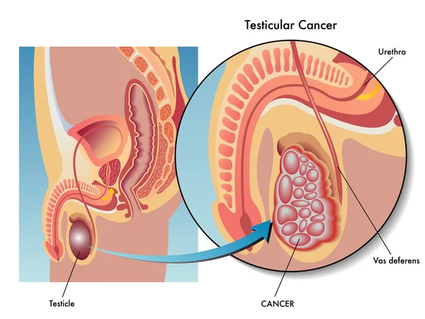
– Breast enlargement (gynecomastia) and/or testicular shrinkage in men

– Increased red blood cell production (erythrocytosis), which may cause a flushed and red appearance

– Elevated cholesterol levels 

What Is Liver Cancer?

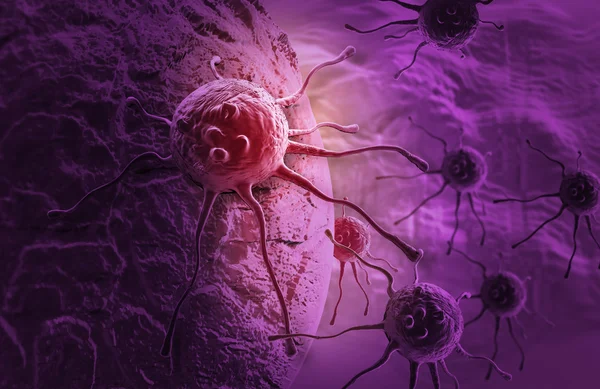
Liver cancer originates in the liver, a vital organ responsible for numerous critical functions. Cancer begins when cells grow uncontrollably. 
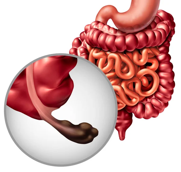
The Liver’s Role

The liver is the largest internal organ, located under the right ribs, beneath the right lung, and divided into two lobes. Primarily composed of hepatocytes, it also contains cells lining the blood vessels and bile ducts. These ducts transport bile from the liver to the gallbladder or intestines. The liver performs essential functions such as:

– Processing and storing nutrients from the intestine; some nutrients must be metabolized in the liver for energy use or tissue repair.

– Producing clotting factors to prevent excessive bleeding from injuries.

– Secreting bile to aid in nutrient (particularly fat) absorption.

– Detoxifying the blood by breaking down alcohol, drugs, and waste products, which are then eliminated through urine and feces.

Various cell types in the liver can lead to different malignant (cancerous) and benign (non-cancerous) tumors, each with its own causes, treatments, and outcomes.

Primary Liver Cancer Types

Hepatocellular Carcinoma (HCC)

Hepatocellular carcinoma is the most prevalent liver cancer in adults, with two growth patterns:

1. A single tumor that enlarges and spreads only in advanced stages.

2. Multiple small cancer nodules throughout the liver, common in those with cirrhosis.

Subtypes of HCC generally do not impact treatment or prognosis, except for fibrolamellar carcinoma. This rare type, often affecting women under 35 without preexisting liver disease, tends to have a better outlook.

Intrahepatic Cholangiocarcinoma

Accounting for 10% to 20% of liver cancers, these originate in the bile ducts within the liver. Although primarily focusing on hepatocellular carcinoma, treatments often overlap for cholangiocarcinomas.

Angiosarcoma and Hemangiosarcoma

These rare cancers develop in cells lining the liver’s blood vessels, often linked to chemical exposures such as vinyl chloride, thorium dioxide, arsenic, or hereditary conditions like hemochromatosis. They usually spread rapidly, making surgical removal difficult. Chemotherapy and radiation can slow progression, though treatment is challenging.

Hepatoblastoma

Primarily affecting children under four, hepatoblastoma resembles fetal liver cells. Successful treatment often involves surgery and chemotherapy, though prognosis worsens if the cancer spreads beyond the liver.

Secondary Liver Cancer

When cancer is discovered in the liver, it often originates elsewhere, termed secondary liver cancer. These metastatic tumors are named and treated based on their primary site. For instance, lung cancer that spreads to the liver is treated as lung cancer, not liver cancer. Secondary liver tumors are more common than primary in many Western countries.

Benign Liver Tumors

Although benign tumors don’t invade nearby tissues or metastasize, they can grow large enough to cause issues. Surgical removal can resolve them.

Hemangioma

The most frequent benign liver tumor stems from blood vessels, usually symptomless but sometimes requiring surgical intervention if bleeding occurs.

Hepatic Adenoma

These originate from hepatocytes and are typically asymptomatic but may cause abdominal pain or bleeding. Due to the risk of rupture and potential malignancy, surgical removal is often recommended. Factors increasing risk include use of certain medications like birth control pills and anabolic steroids, with tumors sometimes shrinking upon discontinuation of these drugs.

Focal Nodular Hyperplasia (FNH)

FNH consists of various cell types and is benign, though it may cause symptoms. Distinguishing FNH from malignant tumors can be challenging, leading to surgical removal for diagnostic clarity. Both hepatic adenomas and FNH are more prevalent in women. 

Stages of Liver Cancer

When a liver cancer diagnosis is made, doctors determine the extent of cancer spread, known as staging. This process is crucial for assessing the cancer’s severity and planning the best treatment strategy. The stage also helps in understanding survival statistics.

Liver cancer stages range from I to IV, with lower numbers indicating less spread. A higher stage, like IV, means more significant spread. Despite individual differences, cancers at the same stage typically have similar treatment approaches and prognosis.

Determining the Stage

Several staging systems exist for liver cancer, but the AJCC (American Joint Committee on Cancer) TNM system is most commonly used in the U.S. It is based on:

– Tumor Size and Extent (T): Assessing the size, number of tumors, and their invasion into nearby structures.

– Lymph Node Involvement (N): Checking if cancer has reached nearby lymph nodes.

– Distant Metastasis (M): Determining if cancer has spread to distant organs like bones or lungs.

Staging Details

Each T, N, and M category is further detailed by numbers or letters, with higher numbers indicating more advanced cancer. This information is combined into stage grouping, leading to an overall stage classification.

– Stage IA: – A single tumor ≤2 cm not invading blood vessels.

– Stage IB: – A single tumor >2 cm without blood vessel invasion.

– Stage II: – A single tumor >2 cm with blood vessel invasion or multiple tumors ≤5 cm.

– Stage IIIA: – Multiple tumors, at least one >5 cm.

– Stage IIIB: – Tumor invasions into major liver veins.

– Stage IVA: – Tumor that has spread to nearby lymph nodes.

– Stage IVB: – Tumor that has metastasized to organs like the bones or lungs.

Other Staging Systems

Due to liver damage often accompanying liver cancer, multiple staging systems account for this complexity, such as:

– Barcelona Clinic Liver Cancer (BCLC) system

– Cancer of the Liver Italian Program (CLIP) system

– Okuda system

These vary globally, with no universal standard. Staging systems like these consider liver function assessment, crucial for treatment decisions.

Child-Pugh Score

The Child-Pugh score assesses liver function, especially in cirrhosis, incorporating factors like:

– Blood bilirubin and albumin levels

– Prothrombin time

– Presence of abdominal fluid (ascites)

– Impact on brain function

Liver function is classified into three classes (A, B, C) based on normality or severity of abnormalities, impacting treatment suitability.

Liver Cancer Classification

Doctors often classify liver cancers based on resectability:

– Resectable or Transplantable Cancers: Can be fully removed surgically; typically early-stage cancers without significant liver damage.

– Unresectable Cancers: Cannot be completely removed due to size, spread, or proximity to vital structures.

– Inoperable Cancer with Local Disease: The patient’s health doesn’t permit surgery despite the cancer’s resectability potential.

– Advanced (Metastatic) Cancers: Spread to distant organs; generally not operable.

If you have questions about the stage of your cancer or the staging system used, discuss it with your doctor for clarity. 

Causes of Liver Cancer

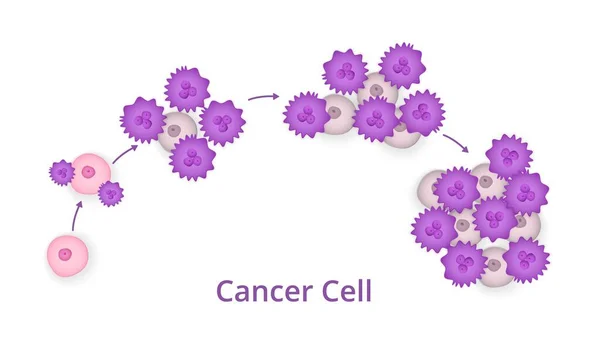
Liver cancer occurs when mutations develop in the DNA of liver cells. DNA contains the instructions that govern every cellular activity in the body. When these mutations alter the instructions, cells may grow uncontrollably, leading to the formation of a tumor composed of cancerous cells. 

In some cases, liver cancer can be attributed to known factors like chronic hepatitis infections. However, it can also occur in individuals without any underlying conditions, where the cause remains unclear.

Risk Factors

Several factors can increase the risk of developing primary liver cancer:

– Chronic Hepatitis Infections: Long-term infections with hepatitis B (HBV) or hepatitis C (HCV) markedly elevate the risk.

– Cirrhosis: This irreversible liver scarring condition raises the likelihood of liver cancer.

– Inherited Liver Diseases: Conditions like hemochromatosis and Wilson’s disease can increase risk.

– Diabetes: Individuals with this condition face a higher risk of liver cancer.

– Nonalcoholic Fatty Liver Disease: Fat buildup in the liver can heighten risk.

– Aflatoxin Exposure: These carcinogenic toxins from molds on poorly stored crops can contaminate food products.

– Excessive Alcohol Use: Long-term, heavy alcohol consumption can lead to irreversible liver damage, increasing cancer risk.

Prevention Strategies

Minimize Cirrhosis Risk

– Moderate Alcohol Consumption: If you drink, limit it to one drink per day for women and two for men.

– Maintain a Healthy Weight: Follow a balanced diet and engage in regular exercise to sustain a healthy body weight.

Prevent Hepatitis Infections

– Hepatitis B Vaccination: Vaccination can significantly reduce the risk of contracting HBV and is available for infants, older adults, and people with weakened immune systems.

– Hepatitis C Precautions: Although vaccines for hepatitis C are unavailable, you can lower the risk by:

  – Knowing the health status of any sexual partners and using protection if necessary.

  – Avoiding IV drug use. If unavoidable, use only sterile needles and avoid sharing.

  – Choosing reputable shops for piercings or tattoos where strict sterilization procedures are followed.

Treat Hepatitis B or C Infections

Effective treatments are available for hepatitis B and C, which can lower the risk of liver cancer.

Liver Cancer Screening

Screening for liver cancer in the general population is not universally recommended due to a lack of evidence that it reduces mortality. However, individuals with increased risk factors, such as those with hepatitis B or C or cirrhosis, might consider screening. Discuss the benefits and potential drawbacks with your doctor to determine if screening, typically involving a blood test and abdominal ultrasound every six months, is appropriate for you. 

Read more

Blood cancer – leukaemia, lymphoma, and myeloma

Blood cancer poses a threat to a vital component of our existence—blood cells. These cells are crucial in providing energy, combating infections, and preventing excessive bleeding. Thankfully, many effective and safe treatment options are available today. Leukaemia, lymphoma and myeloma are some of the most common types of blood cancer.

What is blood cancer?

Blood cancer alters the production and function of blood cells. Typically originating in the bone marrow, where stem cells mature into red and white blood cells and platelets, the disease results in the proliferation of abnormal cells that outnumber healthy ones. This can lead to a cascade of health issues. Advances in treatment have allowed more individuals to live longer with blood cancer.

Are blood cancers serious?

While blood cancers are serious, they are not the most lethal compared to other cancer types. They account for about 10% of all cancer diagnoses in the U.S. annually and roughly 3% of cancer-related deaths. Data from the National Cancer Institute indicates a steady decline in fatalities from blood cancer.

What are survival rates for blood cancer?

Survival statistics are based on averages and may vary by individual. Five-year survival rates help estimate a patient’s health trajectory after diagnosis. Many with blood cancer now have survival expectations similar to those without the disease.

Types of Blood Cancer

Blood cancers are categorized into three main types, each with subtypes:

Leukemia is common, particularly among children and teenagers. Over the last 40 years, its five-year survival rate has quadrupled. Subtypes include acute and chronic forms like acute lymphoblastic leukemia and chronic myelogenous leukemia.

Lymphoma affects the lymphatic system, including the bone marrow. Its survival rate has doubled in the past four decades, with types such as Hodgkin and non-Hodgkin lymphoma.

Myeloma originates in the bone marrow, affecting plasma cells. Multiple myeloma is the most prevalent form, with over half of patients surviving five years post-diagnosis. Other types include plasmacytoma.

Causes and Symptoms

Causes of Blood Cancer

Blood cancer stems from mutations in the DNA of blood cells, whose specific triggers remain unclear. This results in abnormal cells growing faster and living longer than normal cells, crowding out healthy ones.

Leukemia may be triggered by environmental and genetic factors, potentially linked to radiation and chemical exposure.

Lymphoma involves genetic changes in lymphocytes, possibly influenced by infections or immune deficiencies.

Myeloma is linked to genetic changes in plasma cells, with ongoing research into chromosomal influences.

Common Symptoms of Blood Cancer

Symptoms vary, but some are shared across types:

– Extreme fatigue

– Persistent fever

– Night sweats

– Unusual bleeding or bruising

– Unexplained weight loss

– Frequent infections

– Swollen lymph nodes or organs

– Bone pain

These symptoms can mimic other conditions, so persistent changes should prompt a healthcare consultation.

Diagnosis and Testing

Diagnosis begins with a thorough consultation, possibly followed by various tests. Depending on the suspected cancer type, these may include:

– Complete Blood Count (CBC): Measures blood cell levels.

– Blood Chemistry Test: Evaluates blood substances.

– Imaging Scans (CT, MRI, PET): Visualizes internal structures and activity.

– Bone Marrow Biopsy: Analyzes the proportion of normal and abnormal cells.

– Microscopic Blood Examination: Identifies cell abnormalities.

Treatment Options

Blood cancer treatment is personalized, taking into account the individual’s age, health, specific cancer type, and potential treatment side effects. Common treatments include:

– Chemotherapy: Uses drugs to kill cancer cells.

– Radiation Therapy: Targets cancer cells to halt their growth.

– Immunotherapy: Enhances the immune system’s cancer-fighting ability.

– Targeted Therapy: Focuses on specific genetic mutations in cancer cells.

– CAR T-Cell Therapy: Modifies immune cells for better cancer treatment.

– Stem Cell Transplants: Reintroduce healthy cells after intensive treatment.

Treatment Side Effects

Treatments, especially chemotherapy and radiation, have side effects ranging from flu-like symptoms in CAR T-cell therapy to digestive issues in targeted therapies. Stem cell transplants can involve serious complications.

Prevention and Risk Reduction

While specific prevention strategies are elusive due to uncertain causes, certain risk factors have been identified, such as exposure to radiation, certain chemicals, immune deficiencies, family history, and inherited conditions. Awareness and monitoring can help manage potential risks. 

Read more

Basal Cell Carcinoma

Basal cell carcinoma is a form of skin cancer characterized by the development of a bump, lump, or lesion on the epidermis, the skin’s outermost layer. These growths typically appear on skin areas frequently exposed to sunlight. Successful treatment generally results in a favorable prognosis.

An Overview of Basal Cell Carcinoma

Basal cell carcinoma (BCC) originates in the basal cells found at the bottom of the epidermis. It manifests as a small, sometimes glossy bump or a scaly, flat patch that slowly enlarges over time.

What are basal cells?

Basal cells are tiny cells located in the epidermis, the visible layer of the skin. Their primary function is to generate new skin cells by dividing. As new cells emerge, the older cells are pushed to the skin’s surface, where they are shed.

What are the types of basal cell carcinoma?

There are four primary forms of BCC:

1. Nodular: The most prevalent type, presenting as a round, pimple-like bump with visible blood vessels (telangiectasias).

2. Superficial Spreading: Results in small, shallow lesions that appear lighter than the surrounding skin, often found on the torso, arms, and legs.

3. Sclerosing (Morpheaform): Appears as scar-like lesions that gradually expand, typically on the face, sometimes appearing as a small red dot.

4. Pigmented: A rare type causing darkening of the affected skin area compared to the surrounding skin.

Who is Affected by Basal Cell Carcinoma?

BCC can develop in anyone but is slightly more frequent in men. It commonly affects individuals over 50, particularly those with fair skin and light-colored eyes. A history of BCC increases the risk of developing other nonmelanoma skin cancers.

How common is basal cell carcinoma?

This is the most prevalent cancer type overall, as well as the most common skin cancer, with over 4 million cases diagnosed annually in the United States.

Symptoms and Causes

Identifying Basal Cell Carcinoma

Signs include:

– Bumps, lumps, pimples, scabs, or scaly patches on the skin

– Lumps may be translucent and can range from white to pink, or from brown to blue-black

– May shine more than surrounding skin and show tiny blood vessels

– Lumps could grow slowly, be itchy or painful, and sometimes ulcerate or ooze fluid

Locations Prone to Basal Cell Carcinoma

BCC chiefly appears on parts of the body exposed to the sun, such as the face, scalp, nose, eyelids, legs, ears, and arms.

Causes of Basal Cell Carcinoma

The primary cause is a DNA alteration frequently resulting from excessive exposure to ultraviolet (UV) rays from the sun or tanning beds. Genetic mutations may disrupt DNA, impairing cell replication controls, leading to excessive basal cell growth.

Diagnosis and Tests

Diagnosing Basal Cell Carcinoma

Healthcare providers might suspect BCC based on a lesion’s appearance, confirmed by a physical examination and symptom inquiry. Questions may include the appearance timeframe, changes in size, and previous skin cancer history.

Diagnostic Tests for Basal Cell Carcinoma

Skin Biopsy: Removing and examining a skin sample.

Imaging Tests: Rarely, MRIs or CT scans might be performed if cancer spread is suspected.

The diagnosis stage is determined based on the lesion size, growth, and any spread to lymph nodes or other body parts.

Management and Treatment

Treatment for Basal Cell Carcinoma

Cancer removal might involve:

– Electrodessication and Curettage: Scraping and burning cancer with an electric needle.

– Surgery: Excision or Mohs surgery to remove the lesion.

– Cryotherapy: Freezing the lump.

– Chemotherapy: Using drugs to destroy cancer cells.

– Photodynamic Therapy (PDT): Applying light-sensitive agents with blue light.

– Laser Therapy: High-energy lasers replace surgical methods.

Treatment choice considers health, age, lesion size, and location.

Consequences of Untreated Basal Cell Carcinoma

Untreated BCC may grow and affect deeper tissues, causing pain, bleeding, and potentially life-threatening complications if it rarely spreads.

Medication for Basal Cell Carcinoma

For advanced cases, the FDA has approved medications like Vismodegib and Sonidegib for patients ineligible for surgery or radiation, bearing potential side effects like muscle cramps and taste changes.

Surgical Treatment Side Effects

Surgery may cause scarring, with minimal risk of bleeding or infection.

Prevention

Reducing Basal Cell Carcinoma Risk

While prevention isn’t always possible, risk can be minimized by:

– Avoiding sun exposure, especially from 10 a.m. to 4 p.m.

– Steering clear of tanning beds.

– Using sunscreen with an SPF of 30 or higher, applied every two hours when outdoors.

– Wearing protective clothing, hats, and sunglasses.

– Conducting monthly skin self-examinations.

– Having an annual dermatologist skin check.

– Taking vitamin B3 (nicotinamide) twice daily to decrease new BCC occurrences.

Read more